Circulatory health is a fundamental pillar for maintaining a good quality of life, especially in a city with the geographical and demographic richness of Cuenca, Ecuador. Over the years, factors such as a sedentary lifestyle, genetic predisposition, hypertension, and dietary habits can compromise the proper blood flow through our arteries and veins. As a vascular surgery specialist, I often receive patients in my office who are concerned about tired legs, prominent varicose veins, or pain when walking that limits their daily activities. Fortunately, modern medicine has extraordinary, non-invasive, and highly accurate diagnostic tools designed to visualize the interior of our blood vessels without the need for exploratory surgeries. Among them, the vascular doppler study has become the initial gold standard for detecting circulatory problems before they turn into serious conditions such as thrombosis or chronic venous ulcers.
The vascular doppler is an ultrasound diagnostic medical examination that evaluates blood circulation in the body’s arteries and veins. Using high-frequency sound waves, this painless procedure allows real-time visualization of the direction, blood flow velocity, and potential obstructions within the circulatory system.
What is a vascular doppler?
As a vascular surgeon, I usually explain to my patients in Cuenca that a vascular doppler study is like having an open window to observe the behavior of blood inside our vital conduits. This technology combines conventional ultrasound—which shows the anatomical structure of the surrounding vessels and tissues—with the Doppler effect, a physical principle that measures changes in the frequency of sound waves as they bounce off moving red blood cells.
There are mainly two modalities that we apply depending on clinical needs: color doppler, which translates flow velocities and directions into color maps (usually red and blue) superimposed on the grayscale image, and spectral doppler, which graphs flow velocity as a function of time using audible and visual sound waves. According to data from the World Health Organization (OMS/WHO) and statistics from the Ministry of Public Health of Ecuador (MSP), cardiovascular and peripheral vascular diseases represent a significant burden of morbidity, highlighting the crucial importance of early, fast, and accessible diagnosis through this type of ultrasound technology.
When is it indicated?
The range of indications for a vascular echo-doppler is broad, as it can be applied to the lower and upper extremities, the neck vessels (carotids), the abdominal aorta, and the renal vessels. In my daily practice in Cuenca, Ecuador, I frequently indicate it under the following clinical circumstances:
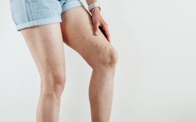
- Chronic venous insufficiency: Presence of visible varicose veins, a feeling of heaviness, burning, swelling (edema), and skin color changes in the legs.
- Deep vein thrombosis (DVT): Suspicion of blood clots in the deep veins, manifested by sudden pain, unilateral swelling, and redness.
- Peripheral artery disease (PAD): Patients reporting calf pain when walking (intermittent claudication) that disappears with rest.
- Cerebrovascular risk evaluation: Detection of atheromatous plaques, stenosis, or narrowing in the carotid and vertebral arteries that could trigger a stroke (CVA).
- Pre- and post-operative monitoring: Planning for varicose vein surgery, peripheral bypasses, or follow-up of arteriovenous fistulas in patients with renal failure.
- Vascular trauma: Evaluation of damage to arteries or veins after physical injuries or accidents.
To learn more about how we approach these venous pathologies, I invite you to discover more about my specialized services in varicose vein treatment in Cuenca, where precise diagnosis with doppler makes all the difference in therapeutic success.
Step-by-step process or treatment
One of the great advantages of vascular doppler is that it is an outpatient, safe, ionizing radiation-free, and completely painless procedure. Below, I detail the step-by-step process of how we perform this study in the office:
- Initial preparation: The patient is received in the examination area. Generally, no fasting or complex preparations are required, although if the study focuses on the aorta or renal arteries, patients may be advised to arrive on an empty stomach to avoid interference from intestinal gases. The patient is asked to remove clothing and accessories from the area to be examined.
- Positioning: The patient lies comfortably on the medical examination table. Depending on whether we are evaluating the arterial or venous system of the legs, carotids, or arms, they will be asked to adopt a supine position (face up), prone position (face down), or even standing up to assess venous valve sufficiency under the effect of gravity.
- Application of conductive gel: A special aqueous gel is applied to the skin along the path of the blood vessels to be studied. This gel eliminates air pockets between the transducer and the skin, allowing optimal transmission of ultrasonic waves.
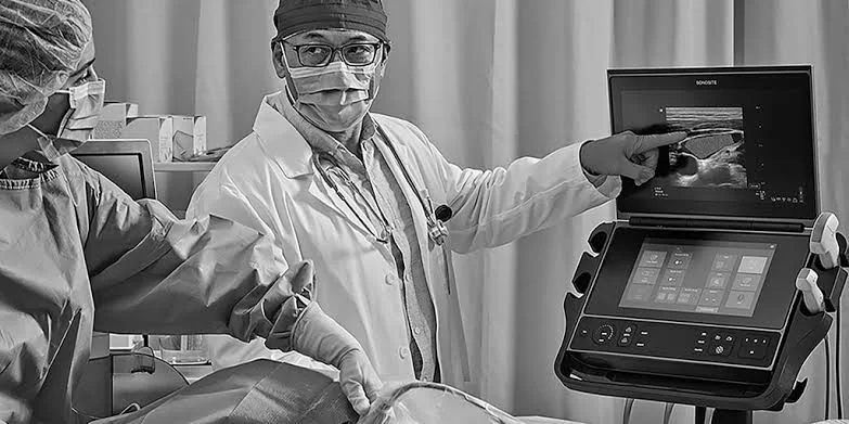
- Ultrasound and doppler exploration: The specialist gently slides the transducer (probe) over the gelled area. The equipment emits sound waves and processes the reflected echoes, displaying real-time images on the screen and emitting the characteristic sound of blood flow (‘whoosh-whoosh’).
- Compression and flow augmentation maneuvers: During the venous exploration, it is common for the physician to perform gentle hand compressions on the calf or thigh to assess valve competence. In arterial studies, segmental pressure measurements can be taken at different points of the extremity.
- Completion and delivery of results: Excess gel is wiped from the patient’s skin. The study takes approximately 20 to 45 minutes depending on the complexity and number of extremities or vascular territories evaluated. Afterward, I proceed to interpret the findings and write the detailed medical report.
Recovery and care
Given that vascular echo-doppler is neither a surgical nor an invasive procedure, recovery is immediate. There are no physical restrictions, medication use, or mandatory rest periods.
Even if the patient comes in for acute pain or suspected thrombosis, they can resume their daily routines, work activities, and walk normally immediately after the test is finished. No wound healing or special subsequent care is required, making this tool an extremely friendly diagnostic resource for people of all ages.
Comparative table: Vascular Echo Doppler vs. Computed Tomography Angiography (CTA)
To help you better understand why doppler is the first-line diagnostic choice compared to other more complex studies, I present the following comparative table based on vascular surgery clinical guidelines:
| Feature | Vascular Echo Doppler | Computed Tomography Angiography (CTA) |
|---|---|---|
| Level of Invasiveness | Completely non-invasive | Minimally invasive (requires an intravenous line) |
| Use of Radiation | Does not use radiation | Uses ionizing radiation |
| Contrast Media | No contrast required | Requires iodinated contrast (renal/allergic risk) |
| Functional Evaluation | Excellent (measures velocity and direction in real time) | Excellent 3D anatomical visualization |
To learn more about how we integrate these technological advancements and comprehensive circulatory health care, I suggest visiting the section on vascular surgery in Cuenca on my official website.
Frequently Asked Questions
Is it painful?
No, the examination is absolutely painless. The only things the patient will experience are a cold sensation due to the conductive gel and slight pressure when the transducer slides over the skin or the doctor compresses the tissues to evaluate the veins.
How long does recovery take?
No recovery time is required. Being a diagnostic ultrasound without punctures or cuts, the patient can get off the examination table and continue with their normal routine immediately.
Does health insurance cover it in Ecuador?
Yes, the vast majority of prepaid medicine policies and private insurance in Ecuador cover vascular echo-doppler when backed by a justified medical order issued by a specialist in vascular surgery or cardiology. In the public system (IESS or Ministry of Health), it is subject to prior evaluation and internal appointment availability.
Is any special preparation required before the study?
Generally, fasting or preparation is not required. However, if the exam is aimed at evaluating the abdominal aorta, renal arteries, or mesenteric vessels, a 6 to 8-hour fast may be requested to reduce the presence of intestinal gases that could obstruct the visualization of deep vessels.
When to consult Dr. Juan Carlos Peralvo?
Timely diagnosis is the difference between simple treatment and the management of complex complications. If you present symptoms such as persistent heaviness in your legs, the appearance of tortuous and bulging veins, nocturnal cramps, unexplained swelling, or pain when walking, you should not postpone your medical check-up. As a specialist in vascular surgery in Cuenca, Ecuador, I am committed to offering you a comprehensive, ethical evaluation backed by cutting-edge technology to take care of your circulatory system. I invite you to schedule an evaluation appointment to design the diagnostic and therapeutic plan that best suits your health needs.